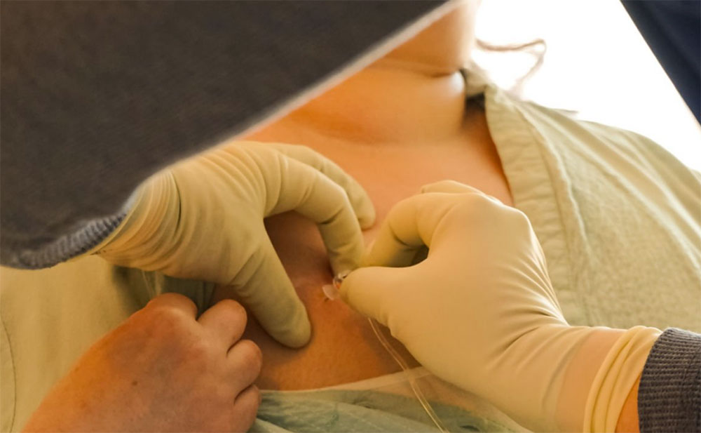
After an ultrasound or mammography, if imaging reveals a breast lump classified as BI-RADS 4A or above, a biopsy is generally recommended. The doctor will use a needle to collect a tissue sample from the lump. This sample will be analyzed by a pathologist to determine if the lump is benign or malignant. Needle biopsy of the breast is very convenient and safe; there is no evidence that undergoing a needle biopsy will cause the malignant tumor to spread or turn a benign tumor into a malignant one.
Fine Needle Aspiration/ Core Needle Biopsy
Fine needle aspiration (FNA) uses a thin needle, similar to those used for drawing blood, to extract only fluids and cells for analysis. It can indicate if the cell is likely benign or malignant, but cytology cannot lead to 100% conclusive results.
Core needle biopsy uses a special trucut needle to extract a strip of tissue, allowing direct observation of tissue structure under a microscope to determine if the tissue is benign, or if malignant, whether it is breast cancer.
Nowadays, cytology via fine needle is rarely performed; most breast biopsies are done by core needle, as it extracts more tissue, gives more accurate results, and helps doctors distinguish tissue types and whether there is invasiveness, which aids in formulating treatment plans.
Vacuum-Assisted Biopsy
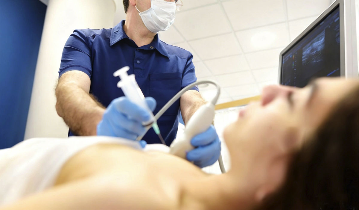
Vacuum-Assisted Biopsy (VAB) is one of the sampling methods. Guided by X-ray or ultrasound, the biopsy needle is directed to the lesion, and vacuum suction helps draw the tissue into the needle. This method collects more breast tissue in one attempt than regular needle biopsy and is useful for sampling X-ray detected lesions, such as microcalcifications. For small benign tumors, VAB can even completely remove the lump. However, not everyone is suited for VAB; its use depends on the type and specific situation, so whether it is suitable should be discussed with the attending doctor.
Imaging-Guided Procedures
Our imaging-guided procedures, including ultrasound-, mammography-, and MRI-guided techniques, ensure accuracy in locating and sampling lesions. Performed by radiologists, these minimally invasive procedures are designed to maximize precision and patient comfort.