Clinical examinations to check for any lumps or abnormalities in the breasts, as well as enlarged lymph nodes in the armpits and neck.
If a lump is found in the breast and feels smooth on the surface, is highly mobile, and has a soft texture when palpated, it is usually benign. Conversely, if the lump has irregular borders, lacks mobility, feels as though it is attached to the breast tissue, or is hard in texture, it may be malignant. However, palpation alone cannot determine whether a breast lump is benign or malignant.
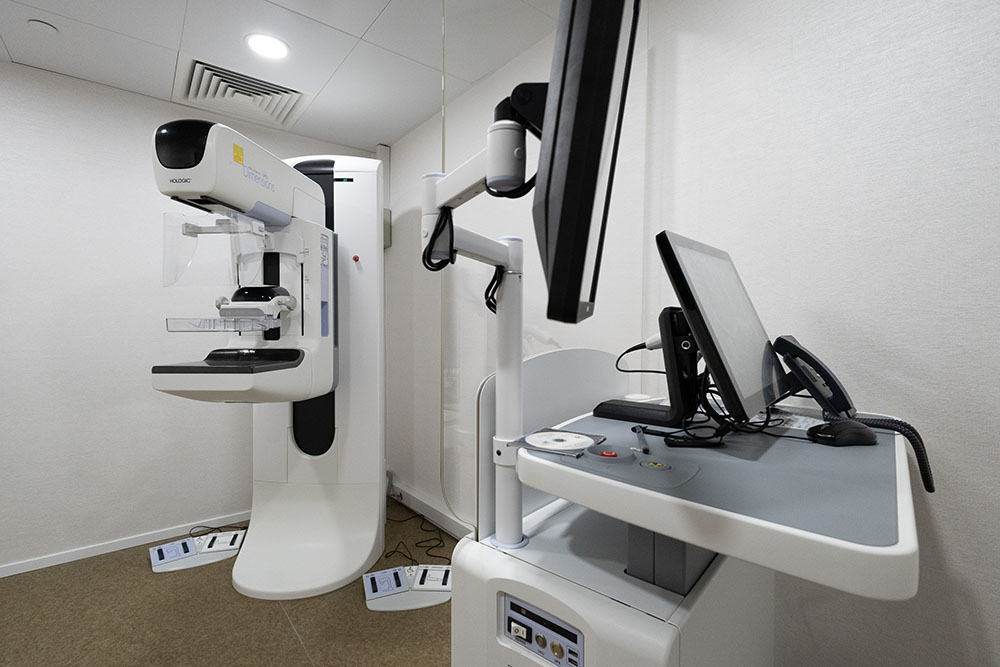
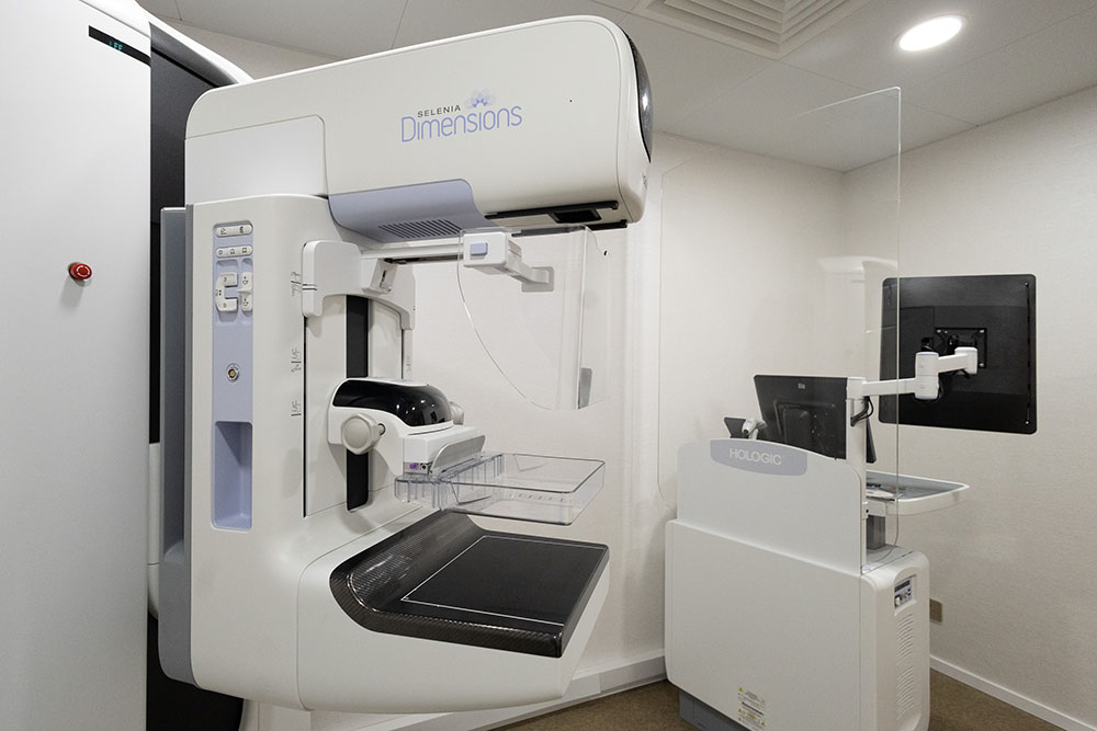
Mammography (2D/3D)
During a mammography examination, you will be required to undress from the waist up and stand in front of the X-ray machine. The technician will assist you in positioning your breast on the platform of the X-ray cassette. A plastic compression plate will gently but firmly press your breast from top to bottom and side to side, allowing the X-ray to capture clear images of the breast tissue. A slight discomfort during compression is common. Two images will be taken of each breast – one from above and one from the side – and the process will then be complete. Studies have shown that mammography can reduce breast cancer mortality by 25% to 30% and remains the most effective method for detecting early-stage breast cancer.
Traditional mammography produces 2D images, in which some areas of dense breast tissue may be obscured. In contrast, 3D mammography – also known as tomosynthesis – captures multiple low-dose images from different angles, each just 1 mm thick, providing a clearer view of breast tissue. This technology improves cancer detection rates, reduces the need for additional imaging, and keeps radiation exposure at the same level as standard 2D mammography.
Mammography is a safe imaging procedure and generally has no side effects. However, women who are pregnant, suspect they may be pregnant, planning pregnancy, or breastfeeding should inform healthcare staff before the examination. Women aged 40 and above are advised to have a mammogram every two years to detect tiny calcifications, but if a lump is palpable, an ultrasound examination should be performed instead.
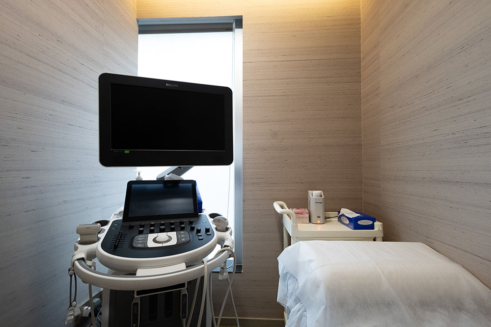
Breast Ultrasound
Breast ultrasound scanning works by transmitting high-frequency sound waves into the breast tissue. The echoes that bounce back are then converted into real-time images, allowing doctors to observe the condition of the breast. Ultrasound is particularly useful for examining cysts or solid lumps, such as fibroadenomas.
During an ultrasound examination, if no dark shadow appears on the image, the findings often indicate benign breast hyperplasia (fibrocystic changes). If a dark area is observed, and the ultrasound image shows a flat shape with well-defined borders, a clear capsule, and uniform internal echoes, it generally suggests a benign lesion.
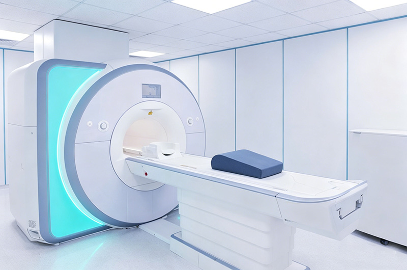
MRI Breast Imaging *
Breast magnetic resonance imaging (MRI) uses magnetic fields to generate detailed images of breast tissue. It is commonly used for women who have already been diagnosed with breast cancer to evaluate the extent of the disease. Additionally, if an ultrasound reveals multiple benign-looking lesions, doctors may recommend an MRI to further assess the possible spread or impact of cancer. MRI is also suitable for high-risk women, such as those under the age of 40 who carry inherited breast cancer genes, allowing for regular monitoring and early detection. Furthermore, MRI can be used to examine breast implants in women who have undergone augmentation surgery.
*Remarks : Referral to a hospital or imaging center for examination based on the patient’s condition.